Phenom XL G2 Desktop SEM applications
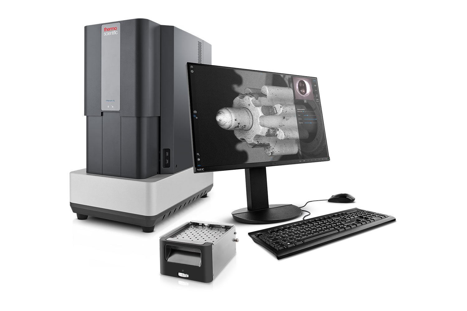
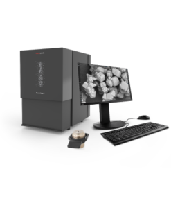
The next-generation Thermo Scientific Phenom XL G2 Desktop Scanning Electron Microscope (SEM) automates your quality control process, providing accurate, reproducible results while freeing up time for value-added work.
The Phenom XL G2 Desktop SEM allow you to:
- Obtain the quality information you need to discover failures early and rapidly adjust your production process when needed.
- Automate quality control to process a high volume of samples with fewer chances of human error.
- Get up to speed quickly with an all-new, easy-to-learn interface ideal for a wide range of applications.
The Phenom XL G2 Desktop SEM features full-screen images and an average time-to image of 60 seconds. The unique CeB6 electron source offers a long lifetime with less maintenance. The small form factor requires little lab space, allowing you to place the microscope exactly where you need it.
Phenom XL G2 Argon-Compatible Desktop SEM
The Thermo Scientific Phenom XL G2 Argon-Compatible Desktop Scanning Electron Microscope (SEM) automates your workflows while providing a stable, non-reactive environment for your reactive sample research.
The Phenom XL G2 Argon-Compatible Desktop SEM offers your research:
- A non-reactive environment to conduct reactive sample research on samples such as solid state lithium batteries
- A modality where you can have sample preparation and SEM/EDX analysis in the same workspace
- A safer way to interface with highly volatile samples such as solid state lithium, enabling the next generation of longer-lasting, eco-friendly batteries
The Phenom XL G2 Argon-Compatible Desktop SEM boasts the same 60-second time to image as the Phenom XL G2 SEM, while also being able to be placed in an argon glove box, assuring the safe analysis of solid state lithium.
Automation
The Phenom XL G2 Desktop SEM is standardly accessible via the Phenom Programming Interface (PPI), a powerful method for controlling the Phenom XL G2 Desktop SEM via Python scripting. If the user has a SEM workflow with repetitive work to analyze particles, pores, fibers or large SEM images, the instrument can do so automatically.
Long-lifetime CeB₆ source
The long-lifetime CeB₆ (cerium hexaboride) source has several advantages. First is the high brightness it provides compared to tungsten, making it much easier for many users to obtain high quality images with many details. Secondly, the lifetime of the source is very long and maintenance can be scheduled.
Eucentric sample holder
In many SEM applications, a user can gain more insight into sample properties if the sample can be tilted and rotated. The optional eucentric sample holder enables eucentric tilt and rotation, making research and analysis faster and more accurate.
Element identification (EID)
The Phenom XL G2 Desktop SEM can be equipped with an optional energy-dispersive X-ray spectroscopy (EDS) detector to obtain more material insights with element identification via X-ray analysis.
Step-by-step data collection
The dedicated software package elemental identification software package (EID) is used to control the fully integrated EDS detector. The intuitive step-by-step process within the EID software helps the user to collect all X-ray results in an organized and structured way.
SEM/EDX preparation and analysis in the same workspace
When placed in an argon glove box, this desktop SEM setup enables your research on solid state lithium batteries since it decreases the risk of sample volatility or sample degradation due to lithium oxidation.
Built-in assurance
This desktop SEM helps you maintain sample integrity by eliminating the need to move your samples to different instruments, saving time and resources.
Fostering the goal of a safe lithium battery
This solution allows you to research solid state lithium batteries safely, enabling the next generation of longer-lasting, eco-friendly, more versatile lithium batteries.
Dry room compatibility
This system has been tested and approved to work in dry room environments with dew temperatures as low as -65 0C.
Process control using electron microscopy
Modern industry demands high throughput with superior quality, a balance that is maintained through robust process control. SEM and TEM tools with dedicated automation software provide rapid, multi-scale information for process monitoring and improvement.
Quality control and failure analysis
Quality control and assurance are essential in modern industry. We offer a range of EM and spectroscopy tools for multi-scale and multi-modal analysis of defects, allowing you to make reliable and informed decisions for process control and improvement.
Fundamental Materials Research
Novel materials are investigated at increasingly smaller scales for maximum control of their physical and chemical properties. Electron microscopy provides researchers with key insight into a wide variety of material characteristics at the micro- to nano-scale.
Technical Cleanliness
More than ever, modern manufacturing necessitates reliable, quality components. With scanning electron microscopy, parts cleanliness analysis can be brought inhouse, providing you with a broad range of analytical data and shortening your production cycle.
EDS Elemental Analysis
Thermo Scientific Phenom Elemental Mapping Software provides fast and reliable information on the distribution of chemical elements within a sample.
3D EDS Tomography
Modern materials research is increasingly reliant on nanoscale analysis in three dimensions. 3D characterization, including compositional data for full chemical and structural context, is possible with 3D EM and energy dispersive X-ray spectroscopy.
Atomic-Scale Elemental Mapping with EDS
Atomic-resolution EDS provides unparalleled chemical context for materials analysis by differentiating the elemental identity of individual atoms. When combined with high-resolution TEM, it is possible to observe the precise organization of atoms in a sample.
Multi-scale analysis
Novel materials must be analyzed at ever higher resolution while retaining the larger context of the sample. Multi-scale analysis allows for the correlation of various imaging tools and modalities such as X-ray microCT, DualBeam, Laser PFIB, SEM and TEM.







 VI
VI