Bắn cá - Công ty TNHH trò chơi

Online Support

Tundra Cryo – Transmission Electron Microscopy (TEM)

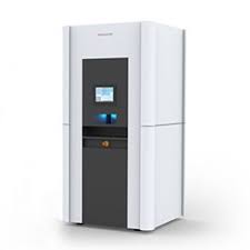
The Thermo Scientific Tundra Cryo-Transmission Electron Microscope (Cryo-TEM) allows you to perform structural analyses on challenging proteins and macromolecules with unprecedented ease of use, at a price point within the reach of many grants and funding sources.

Bringing cryo-electron microscopy to you

Whether you’re an experienced microscopist, or just starting out, the Thermo Scientific Tundra Cryo-Transmission Electron Microscope (Cryo-TEM) is a dedicated structural analysis solution designed to bring cryo-electron microscopy (cryo-EM) to your laboratory at an affordable price point. Simplified data collection and rapid sample loading/unloading make the Tundra Cryo-TEM a powerful tool for answering your most challenging research questions. With the Tundra Cryo-TEM, you can obtain structures at biologically relevant resolutions in-house or reliably prepare high-quality samples for further analysis on Thermo Scientific Krios or Glacios Cryo-TEMs.

Source
  • X-FEG (extreme high-brightness field emission gun)
Accelerating voltage
  • Fixed accelerating voltage of 100 kV
Semi-automated sample loading
  • Cryo-preparation station for contamination free sample exchange
  • Sample transfer device for transfer of single AutoGrids to the microscope, with fixed cryo-box
Lenses
  • High-resolution objective lens optimized for single particle analysis
Stage
  • Computerized specimen cryo-stage
Imaging options
  • High-performance Falcon C Direct Electron Detector for improved resolution capabilities
  • Standard Ceta-F CMOS Camera with dose fractionation, optimized for low-dose imaging
Room size requirements (L x W X H)
  • All in one room dimensions: 4.20 x 4.00 x 2.74 m (13.8 x 13.1 x 8.99 ft)
  • Dimensions with cryo-loading station in neighboring room: 4.00 x 3.80 x 2.74 m (13.1 x 12.5 x 8.99 ft)
Software
  • AI-enabled and automated software solution that provides feedback during data collection and has an open application programming interface

The Tundra Cryo-TEM is equipped with a 100 kV high-brightness field emission gun (X-FEG) that allows for high resolution structure determination at routinely 3-5 Å. The Tundra Cryo-TEM can be installed with either the Falcon C Direct Electron Detector or Thermo Scientific Ceta F CMOS Detector. The Falcon C Direct Electron Detector provides a boost in contrast for single particle cryo-EM, improving resolution and obtaining results faster with less data to maximize your resolution efforts. Tundra Cryo-TEM with the Falcon C Direct Electron Detector makes it possible to efficiently solve protein structures 100 kDa or less.

Single particle cryo-EM analysis

The 100 keV Tundra Cryo-TEM can efficiently validate ligand and protein binding when in fast mode, taking over 10,000 images in as little as a few days’ acquisition time. With the Tundra Cryo-TEM, you can take a closer look at receptors that affect the central nervous system or investigate prokaryotic ribosomes. From Nobel –prize-winning discoveries, like TRPV5, to small potential drug targets, like GABAA receptors, the Tundra Cryo-TEM can help expand your scientific discovery by visually resolving structures and producing 3D reconstructions at ~2–5 Å resolution. When the Tundra Cryo-TEM is configured with a Falcon C Direct Electron Detector, the resolution can be improved, particularly for small and asymmetric protein complexes such as transthyretin (55 kDa).

Molecular structures determined using the Tundra Cryo-TEM

2.1 Å, Apoferritin (~500 kDa)

  • Number of images: 5,418
  • Number of particles: 249,808 (octahedral)
  • Acquisition time: 18 hrs
  • Detector: Falcon C

Drug discovery with the Tundra Cryo-TEM

Cryo-electron microscopy (cryo-EM) is rapidly drawing interest in structure-based drug discovery and design, since it can accurately and rapidly visualize to high resolutions the interactions between drug and receptor of a multitude of samples.

Cryo-TEM sample optimization and connectivity

The Tundra Cryo-TEM can be used to optimize samples, offering efficient sample iteration and optimization. It serves as a valuable screening tool, providing biochemical sample optimization in less time than higher-level cryo-EM instruments. In the example of CAK complex, when using Krios G4 Cryo-TEM, higher resolution reconstruction can be achieved to reveal more structural information.

Room temperature TEM applications

The Tundra Cryo-TEM can be utilized for negative-stain electron microscopy, an easy and cost-effective method for the quality assessment of purified biological specimens at room temperature to quickly assess the stability, homogeneity, and concentration of purified samples. In addition, room temperature sections of resin-embedded cells and tissues or isolated particles of protein complexes and viral assemblies can also be visualized with the Tundra Cryo-TEM.